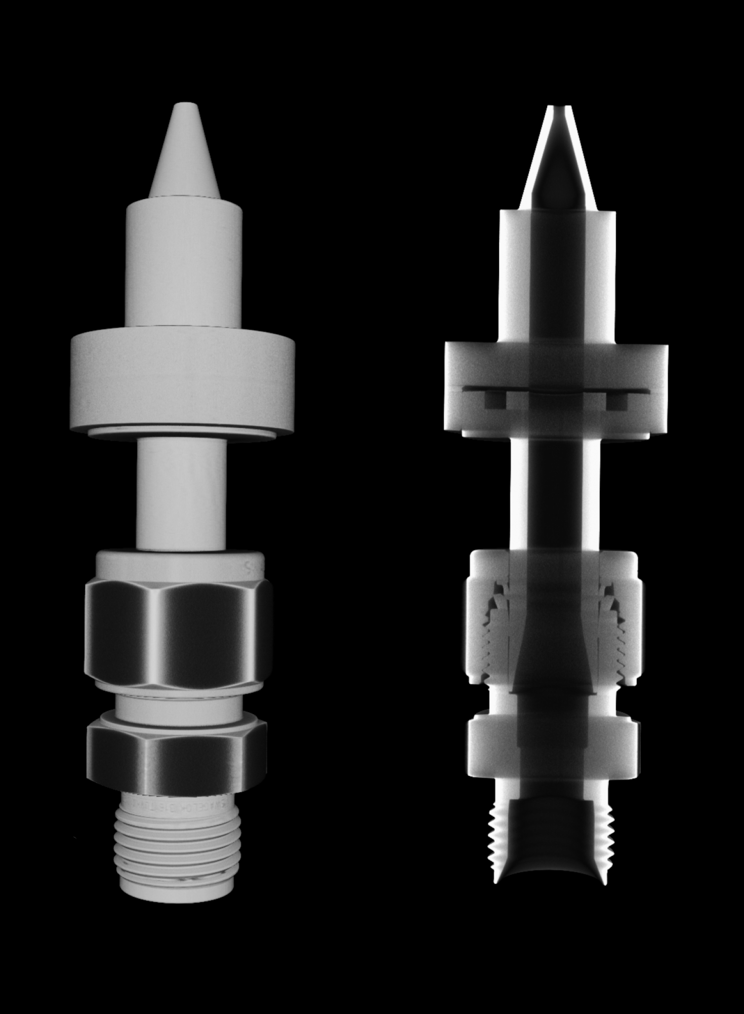
This image shows the internal construction of the "Motive nozzle", a crucial component of the two-phase ejector, which is widely used in refrigeration and air conditioning applications. This motive nozzle is typically a converging-diverging nozzle through which a high-pressure fluid expands, and upon expansion, a very high velocity (often sonic or supersonic speed) is achieved at the motive nozzle outlet. This part, along with other ejector components, is used to improve system performance in HVAC&R systems. This image was produced using the "North Star Imaging X5000 2D Digital Radiography and 3D Computed Tomography", and implementing the vortex scan technique. This image shows the exterior (left) and interior (right) detail geometry for the motive nozzle, which would provide valuable insights into the dimensional variations for manufacturing these components and how these dimensional variations influence the HVAC&R system performance.
Author: Md Muntasir Alam - Energy Transport Research Lab, MechSE
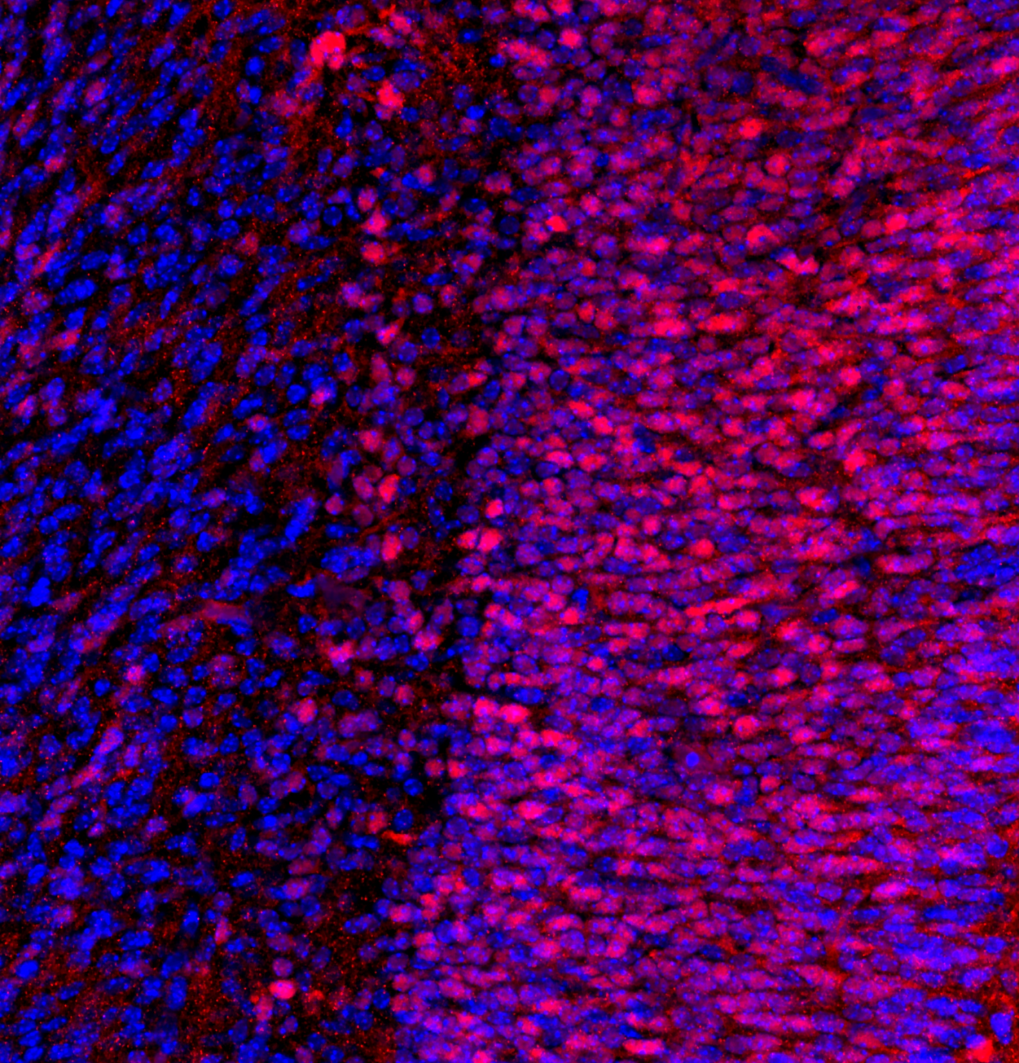
"Representative coronal image from one hemisphere of an E16.5 mouse embryo brain section (25 μm thick) stained for NeuN (red) and Hoechst (blue). NeuN is a neuronal nuclear protein that serves as a marker of post-mitotic neurons, allowing identification of maturing neuronal populations. Hoechst is a fluorescent DNA-binding dye that labels all cell nuclei, providing structural context. Image was acquired using a Zeiss LSM 880 confocal microscope with a 20× objective. We thank Fernando Rigal for his assistance with microscope settings"
Image provided by Rafael J. Gonzalez-Ricon from the Antonson Lab


While the mechanisms driving activation of the innate immune response during viral infection are well understood, the spatial organization remains poorly defined. Using in vitro models of the respiratory tract, we aim to dissect how spatial structure shapes the activation of the innate immune response during influenza A virus infection.
Axioscan image of human bronchial epithelial cells (HBECs). Stained with DAPI (blue) and Phalloidin (green)
Joel Rivera Cardona from the Brooke Lab


This is an image of a gelatin methacrylate model for the perivascular niche of the bone marrow. The perivascular niche, defined by proximity to arteriolar and sinusoidal vessels that comprise endothelial and mesenchymal cells within the bone marrow, regulates hematopoietic stem cell proliferation, differentiation, and quiescence through secreted factors, cell-cell interactions, and local remodeling of the bone marrow.
In this image, we have Hoechst 33342-stained nuclei in blue, murine bone marrow-derived mesenchymal stem cells (mMSCs) stained for podoplanin in cyan, and murine bone marrow-derived endothelial cells (mBMECs) stained for CD31 in green.
Gunnar Thompson from the Harley Lab


Two-photon image deep into the whole brain of a mouse fetus (Gestational Day 16.5) and post-processed with the Spectral Unmixing Module. Shown is the cerebellum structure and labels are for: CD31 (Green), Claudin-5 (red), Hoechst (Blue). The fetus was cleared with an adapted form of a new tissue clearing technique called ADAPT-3D.
Image taken with LSM 980
Image Courtesy: Fernando Rigal from the Antonson Lab

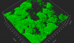
3D reconstruction of Salmonella Typhimurium biofilms in presence of differentially charged micro-nanoplastics (MNP). The study examined how MNP exposure influences bacterial attachment, aggregation, and extracellular polymeric substance (EPS) production- key factors that enhance biofilm resilience in food processing environments. Confocal laser scanning microscopy (CLSM) was used to visualize biofilm architecture and development over time. Understanding these interactions is critical, as enhanced biofilm formation may increase bacterial persistence, antimicrobial resistance, and the risk of contamination, posing serious threats to public health.
LSM 880, stained with FilmTracerFM 1-43 green biofilm cell stain
From Jayita De in the Pratik Banerjee Lab


An unexpected discovery of a bird skeleton within a nest built in a wooden enclosure provides new insights into nesting behavior and material composition. This finding informs the ongoing design of a concrete nest, exploring the intersection of natural instincts and architectural innovation.
Author: Arpit Amarseda


"This SBF-SEM image displays the cell body of a CA1 pyramidal neuron in the hippocampus. Our study aims to investigate mitochondrial morphology and complexity in the mouse hippocampal neurons."
Image taken on Zeiss Sigma VP SBF-SEM
Authors: Kingsley Boateng (IGB Core) and Vipendra Kumar (Nien-Pei Tsai’s
Lab).

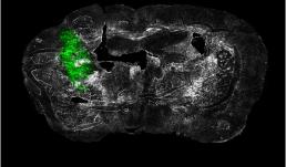
"Ex vivo coronal section of glioma cells, labeled with GFP (green), in a mouse model of glioblastoma (GBM) on day 14 of tumor development."
Image taken on Axioscan.
Author: Urbi Saha - Andrew Smith's Lab


Cryptosporidium parvum is a protozoan parasite with a single host lifecycle composed of asexual (trophozoites and meronts) and sexual (female and male gamonts) replicative stages. This parasite is microscopic in size (~2-6 µm) when compared to its host cells (~25µm). To visualize the developmental stages of the parasite in higher resolution we labeled parasite proteins and utilized antibodies, stains, and SR-SIM. In the leftmost image there are two meronts (in green parasite protein, red parasite stain, blue nuclear stain), on top right corner the merozoites are escaping from the vacuole and on the bottom, there is a mature meront. The center image shows two male gamonts (in red parasite protein, green parasite stain, blue nuclear stain) and two female gamonts (green parasite stain) next to each other. The rightmost image shows mature male gametes escaping from the gamont in an event refer to as “egress”.
Author: Maria Nava - Sumiti Vinayak Lab.

View Gallery
1
/
10