Lightsheet

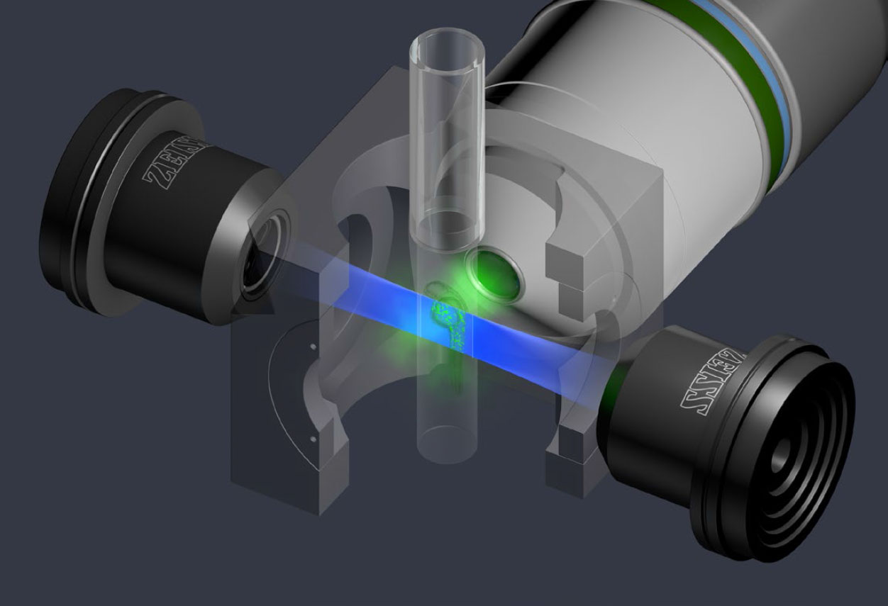
The Lightsheet Z1 system is a live cell and whole organ/scaffold imaging microscope. Compared to conventional fluorescence and confocal microscopy, this is the first system where the illumination axis and the detection axis are separated and placed orthogonal to each other. This system has two SCMOS cameras for simultaneous imaging in two channels and has the following specific features and advantages over fluorescence and confocal microscopes.
Orthogonal Plane Illumination of Excitation and Emission
Reduced Photobleaching and Phototoxicity
No Out of Focus Excitation
Live Cell Culture and Imaging with Temp and CO2 Controls
Large Whole Tissue or Animal Imaging
Lasers: 405, 488, 561 and 633 nm
Dual SCMOS Sensor over 2kx2k Resolution with Large Field of View 1.2x1.2 mm (20x)
Fast Acquisition for Time Lapse (~1000 slices full frame in ~3 min)
Single Side or Dual Side Illumination and Fusion of Multiple Angles
Ideal for Large Optically Cleared Specimens
Lightsheet Sample Preparation Guide
System Principle and Pivot Scan References
Sample Images
Isosurface of Live and Dead Cells in a Scaffold of ~1 cubic mm

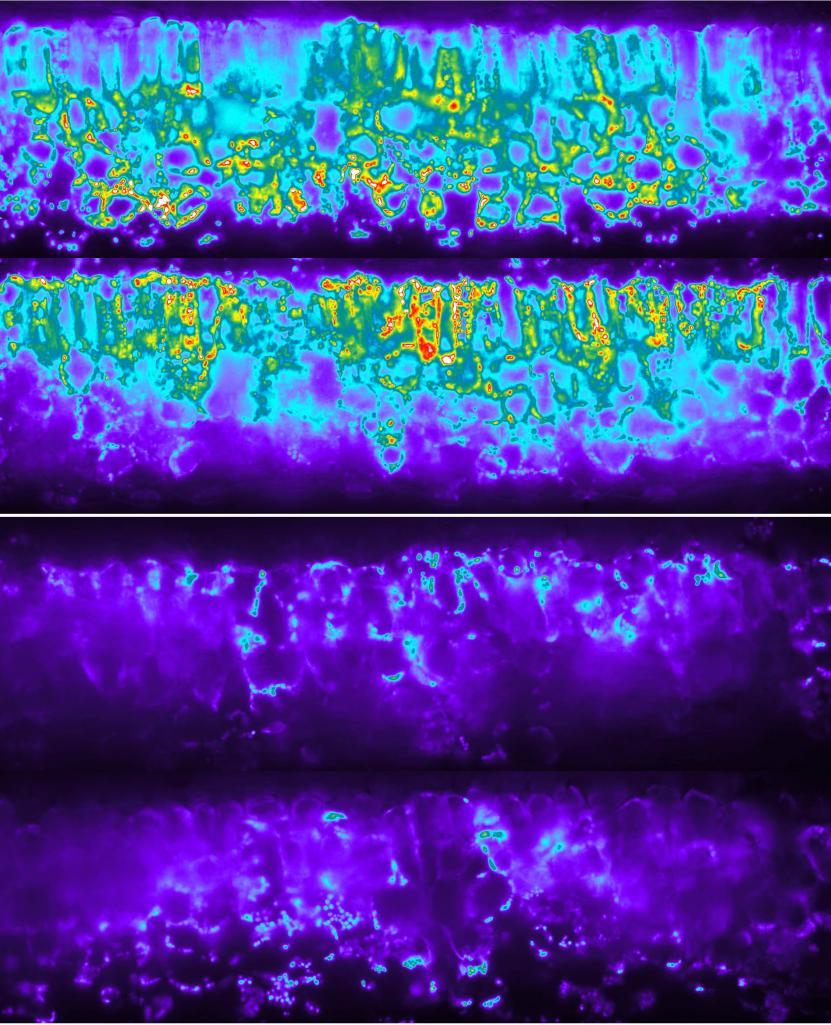
Orthogonal Imaging of Plant Photosynthetic Components

Lightsheet Application and Zen Black Edition Lite Download
Contact Person: Kingsley Boateng, 217-300-1642, kboateng@illinois.edu